Comparative analysis of fixation and embedding techniques for optimized histological preparation of zebrafish.
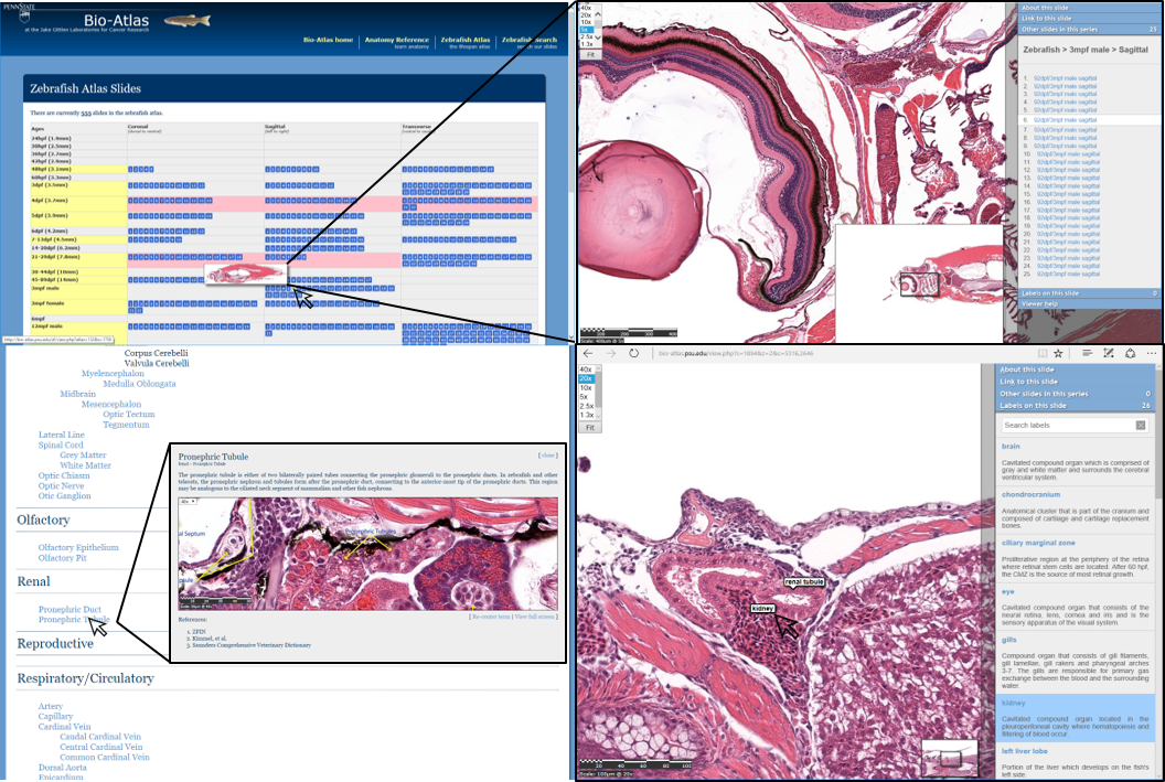
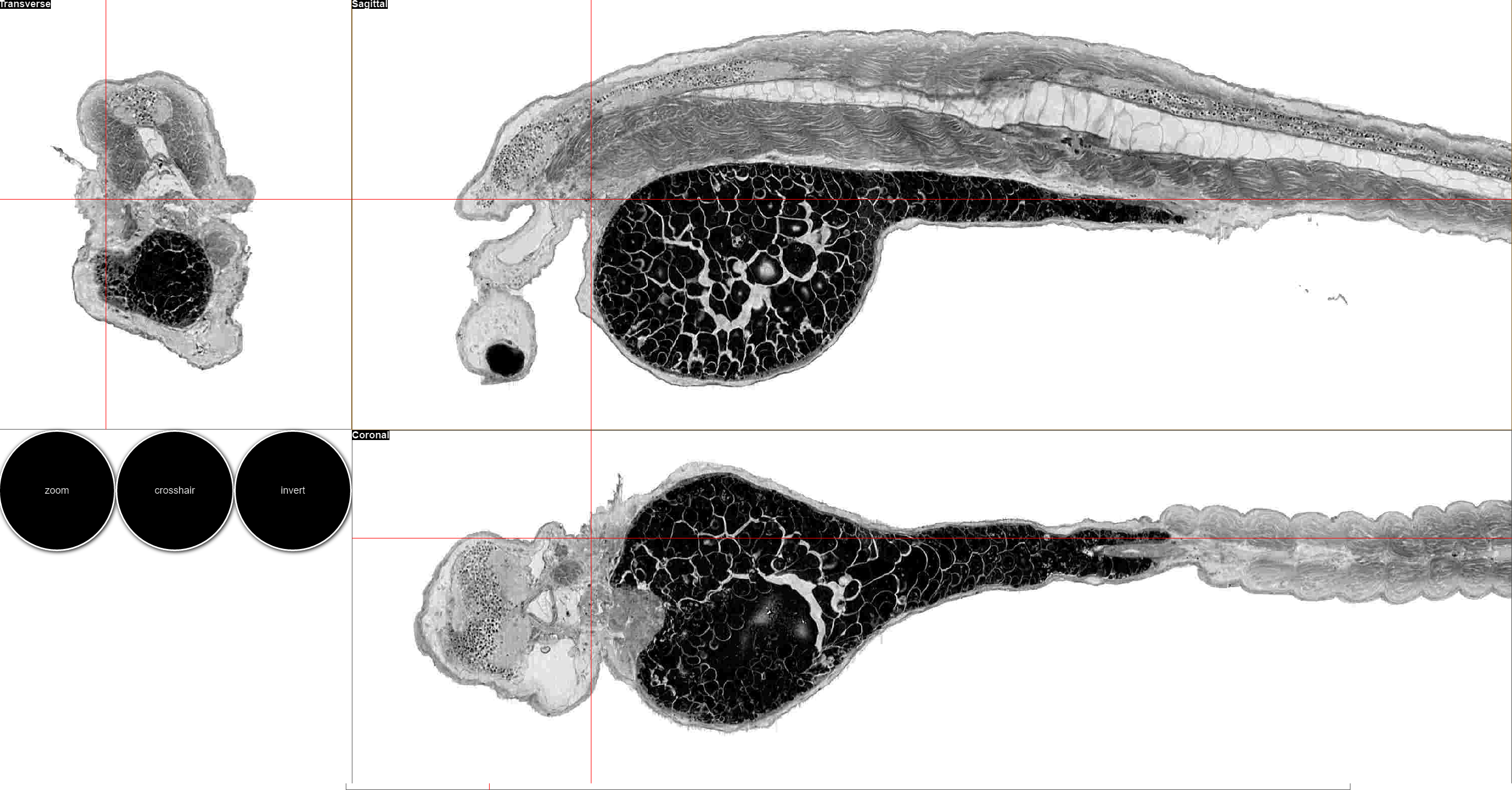
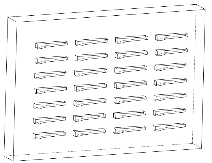
In recognition of the importance of zebrafish as a model organism for studying human disease, we have created zebrafish content for a web-based reference atlas of microanatomy for comparing histology and histopathology between model systems and with humans (bio-atlas.psu.edu). Fixation, decalcification, embedding, and sectioning of zebrafish were optimized to maximize section quality. A comparison of protocols involving six fixatives showed that 10% Neutral Buffered Formalin at 21°C for 24h yielded excellent results. Sectioning of juveniles and adults requires bone decalcification; EDTA at 0.35M produced effective decalcification in 21-day-old juveniles through adults (≥~3Months). To improve section plane consistency in sets of larvae, we have developed new array casting molds based on the outside contours of larvae derived from 3D microCT images. Tissue discontinuity in sections, a common barrier to creating quality sections of zebrafish, was minimized by processing and embedding the formalin-fixed zebrafish tissues in plasticized forms of paraffin wax, and by periodic hydration of the block surface in ice water between sets of sections. Optimal H&E (Hematoxylin and Eosin) staining was achieved through refinement of standard protocols. High quality slide scans produced from glass histology slides were digitally processed to maximize image quality, and experimental replicates posted as full slides as part of this publication. Modifications to tissue processing are still needed to eliminate the need for block surface hydration. The further addition of slide collections from other model systems and 3D tools for visualizing tissue architecture would greatly increase the utility of the digital atlas.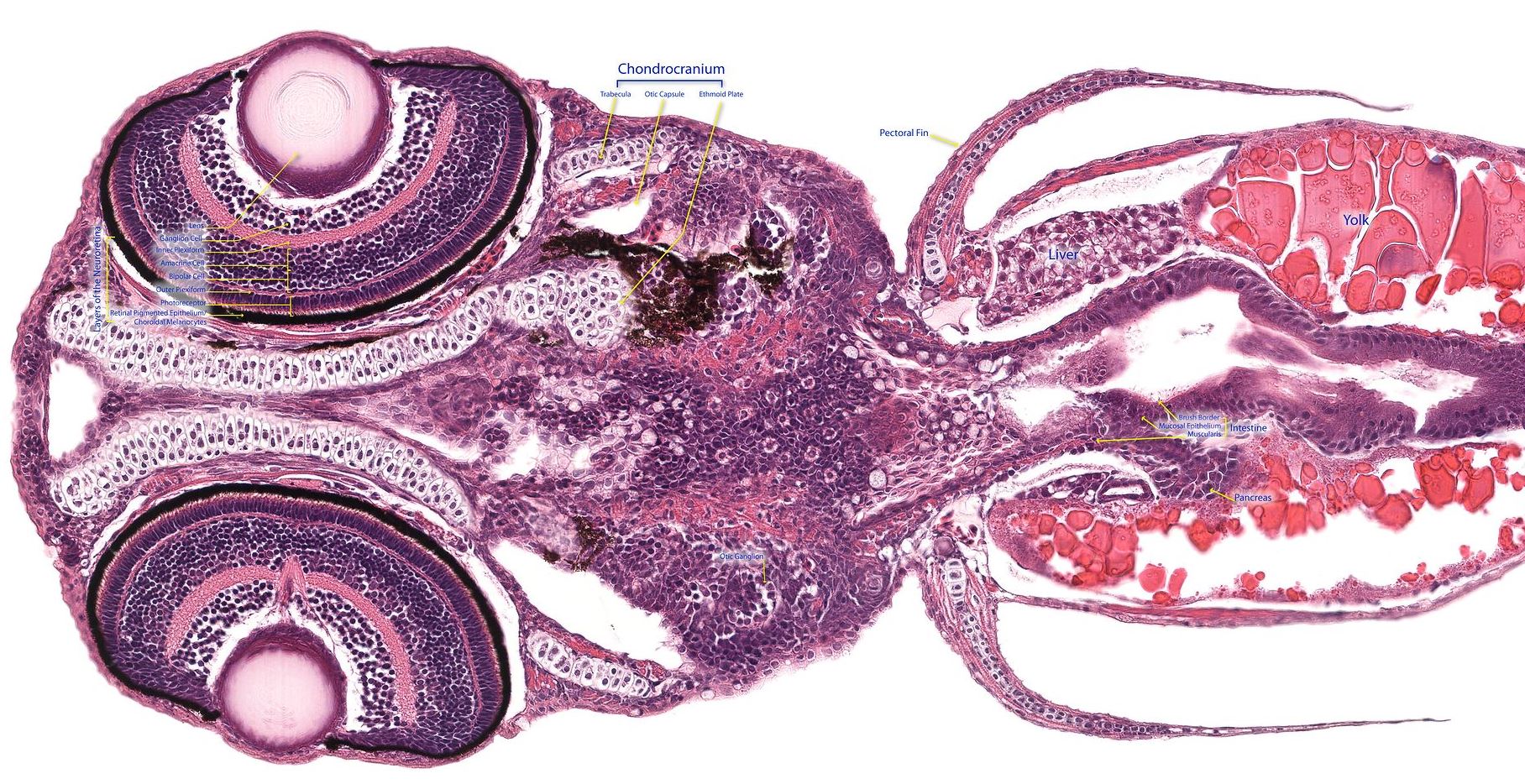
In recognition of the importance of zebrafish as a model organism for studying human disease, we have created zebrafish content for a web-based reference atlas of microanatomy for comparing histology and histopathology between model systems and with humans (bio-atlas.psu.edu). Fixation, decalcification, embedding, and sectioning of zebrafish were optimized to maximize section quality. A comparison of protocols involving six fixatives showed that 10% Neutral Buffered Formalin at 21°C for 24h yielded excellent results. Sectioning of juveniles and adults requires bone decalcification; EDTA at 0.35M produced effective decalcification in 21-day-old juveniles through adults (≥~3Months). To improve section plane consistency in sets of larvae, we have developed new array casting molds based on the outside contours of larvae derived from 3D microCT images. Tissue discontinuity in sections, a common barrier to creating quality sections of zebrafish, was minimized by processing and embedding the formalin-fixed zebrafish tissues in plasticized forms of paraffin wax, and by periodic hydration of the block surface in ice water between sets of sections. Optimal H&E (Hematoxylin and Eosin) staining was achieved through refinement of standard protocols. High quality slide scans produced from glass histology slides were digitally processed to maximize image quality, and experimental replicates posted as full slides as part of this publication. Modifications to tissue processing are still needed to eliminate the need for block surface hydration. The further addition of slide collections from other model systems and 3D tools for visualizing tissue architecture would greatly increase the utility of the digital atlas.

Our understanding of European skin is already partially delineated: in 2005, our lab uncovered one of the most pervasive genetic mutations (SLC24A5) that contribute to the light color of European skin. Europeans also carry a 10 to 20-fold increase in melanoma susceptibility (one of the deadliest forms of skin cancer) when compared to Africans. Surprisingly, East Asians, unlike Europeans, carry a seemingly unusual protection against melanoma: their incidences of disease are similar to those of Africans, despite their relatively light skin color due to yet unknown genetic pathways. To understand this misnomer, the first step is to identify these East Asian-specific genetic mutations.
Our understanding of European skin is already partially delineated: in 2005, our lab uncovered one of the most pervasive genetic mutations (SLC24A5) that contribute to the light color of European skin. Europeans also carry a 10 to 20-fold increase in melanoma susceptibility (one of the deadliest forms of skin cancer) when compared to Africans. Surprisingly, East Asians, unlike Europeans, carry a seemingly unusual protection against melanoma: their incidences of disease are similar to those of Africans, despite their relatively light skin color due to yet unknown genetic pathways. To understand this misnomer, the first step is to identify these East Asian-specific genetic mutations.
Answers to the basic question of how and why gene function is lost in somatic tissues will contribute to our understanding of aging and some forms of human disease, including cancer. Those mutations play a key role in the evolution of killer cancer cells from the originally normal ones of cancer victims, and also the evolution of resistant cancer cells after treatment. The tendency to mutate one’s DNA can be called genetic instability or genomic instability, and the phenotype of elevated mutation rate is called mutator phenotype. In order to discover new vertebrate genes that control mutation, we have used the zebrafish (Danio rerio) to generate mutants that show elevated rates of somatic (body cell) mutation. In this screen, we scored for increased somatic loss of heterozygosity at a marker locus, golden. We expect genetic instability to be caused by deficiencies in any of a number of functions, including chromosome segregation, recombination, and DNA repair. We are studying the characteristics of mutants, including the ability of the mutations to significantly increase cancer susceptibility, and are engaged in the positional cloning of these mutations. Insights gained from these studies will increase our understanding of the molecular forces that drive evolution and may suggest new ways to fight cancer. Since these genomic instability (“gin”) mutants tend to develop cancer, they represent an animal model for human genetic syndromes that predispose to cancer, and may promote the detection of environmental mutagens. This novel approach to the study of genetic instability was sponsored originally by the Jake Gittlen Memorial Golf Tournament, American Cancer Society, and the National Science Foundation.

5dpf Wild Type
4dpf HHT

webview 3

webview 4

webview 5

webview 6

The atlas project was originally focused on the microanatomy of zebrafish and humans, viewable as virtual slides at bio-atlas.psu.edu. The content is being re-organized to have subsections devoted to zebrafish, humans, mice, and other model systems. We are working on ways to present microCT-based 3D histology. We welcome ideas and high-resolution slides and images from the community and are seeking collaborations that would make the resource more valuable.
The Bio-Atlas is organized with subsections devoted to humans, zebrafish, mice, and other model systems. The project was originally focused on the microanatomy of zebrafish. To address the need for a more human orientation, we introduce the Bio-Atlas as a platform for coordination of data between human and other model systems.
Since pathophysiological mechanisms in humans are reflected in tissue architecture, morphological changes in the best models of human disease will involve similar phenotypes as in humans. We welcome ideas and high-resolution 2D and 3D images from the community and are seeking collaborations to expand this resource.